Figure 2.
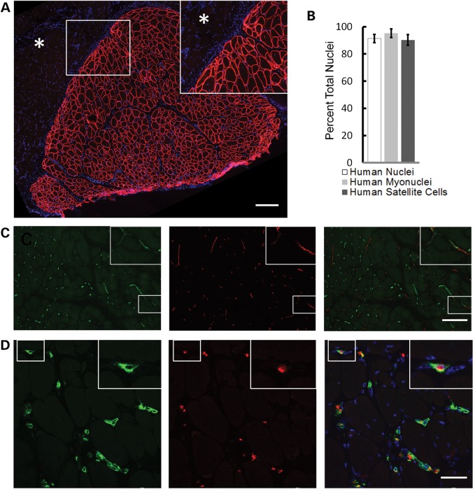
Skeletal muscle xenografts have human myofibers, nuclei and capillary immunoreactivity. (A) Anti-human spectrin antibodies (red) define discrete boundaries (insert) of xenograft (serial section of 1E) from neighboring mouse muscle (asterisk) and uniformly recognize myofiber membrane. Scale bar: 200 µm. (B) Within anti-human spectrin defined xenograft, 91.4 ± 3.4% of all nuclei (DAPI+), 95.2 ± 3.2% of myonuclei and 90.2 ± 4.0% of Pax7+ satellite cells are human as recognized by anti-human lamin A/C, (n = 6). Data are shown as the mean ± SD. (C) Both human (green) and mouse (red) vascular networks were identified with species-specific CD31 antibodies, which appear to anastomose (insert). Scale bar: 200 µm. (D) Human capillaries (green) within the xenograft contain mouse erythroid cells identified with species-specific antibodies (red). Xenografts in these images are all 130 days posttransplantation. Scale bar: 50 µm.

