Figure 1.
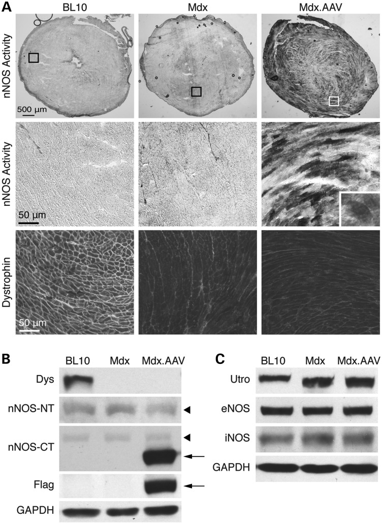
Robust myocardial expression of ΔPDZ nNOS. Aged female mdx mice were infected with a tyrosine mutant AAV-9 vector expressing the ΔPDZ nNOS gene. Animals were euthanized 7 months later for immunostaining and western blot study. (A) Dystrophin immunostaining and nNOS activity staining. Top panel: representative photomicrographs of nNOS activity staining of the full-view heart section. Middle and bottom panels: representative photomicrographs of nNOS activity staining and dystrophin immunostaining of the respective boxed areas in the top panel. Inset: a high-power image showing even distribution of ΔPDZ nNOS in a cardiomyocyte. (B) Immunoblot for dystrophin (Dys), nNOS and Flag. nNOS was detected with an nNOS N-terminal-specific antibody (nNOS-NT) and an nNOS C-terminal-specific antibody (nNOS-CT). Arrowhead: endogenous nNOS; arrow: ΔPDZ nNOS. (C) Immunoblot for utrophin (utro), eNOS and iNOS. GAPDH serves as the loading control for immunoblot.

