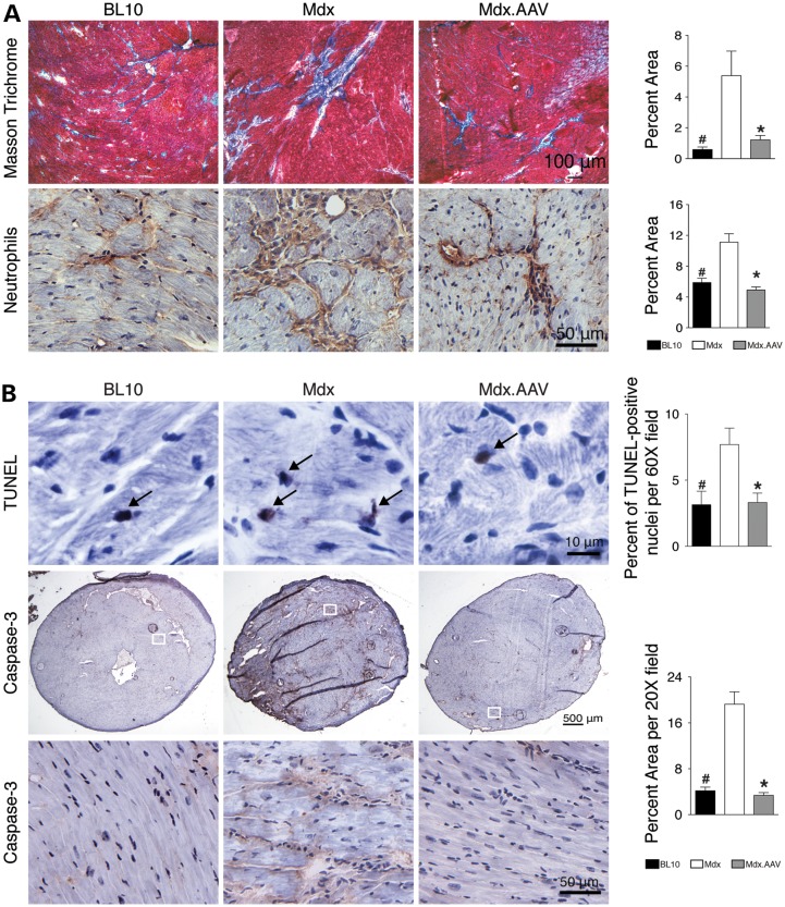
Figure 3.
ΔPDZ nNOS expression reduces myocardial fibrosis, inflammation and apoptosis. (A) Representative photomicrographs of Masson trichrome staining (for fibrosis) and neutrophil immunohistochemical staining. Bar graphs show quantification results. N = 3, 4 and 6 for BL10, untreated and treated mdx, respectively. (B) Representative photomicrographs of TUNEL assay and Caspase-3 immunostaining. Bar graphs represent quantification results. N = 3, 4 and 6 for BL10, untreated and treated mdx, respectively, in TUNEL assay; N = 3, 4 and 6 for BL10, untreated and treated mdx, respectively, in Caspase-3 immunostaining. Asterisk: treated mdx is significantly different from that of untreated mdx; Pound sign: BL10 is significantly different from that of untreated mdx.

