
Figure 3.
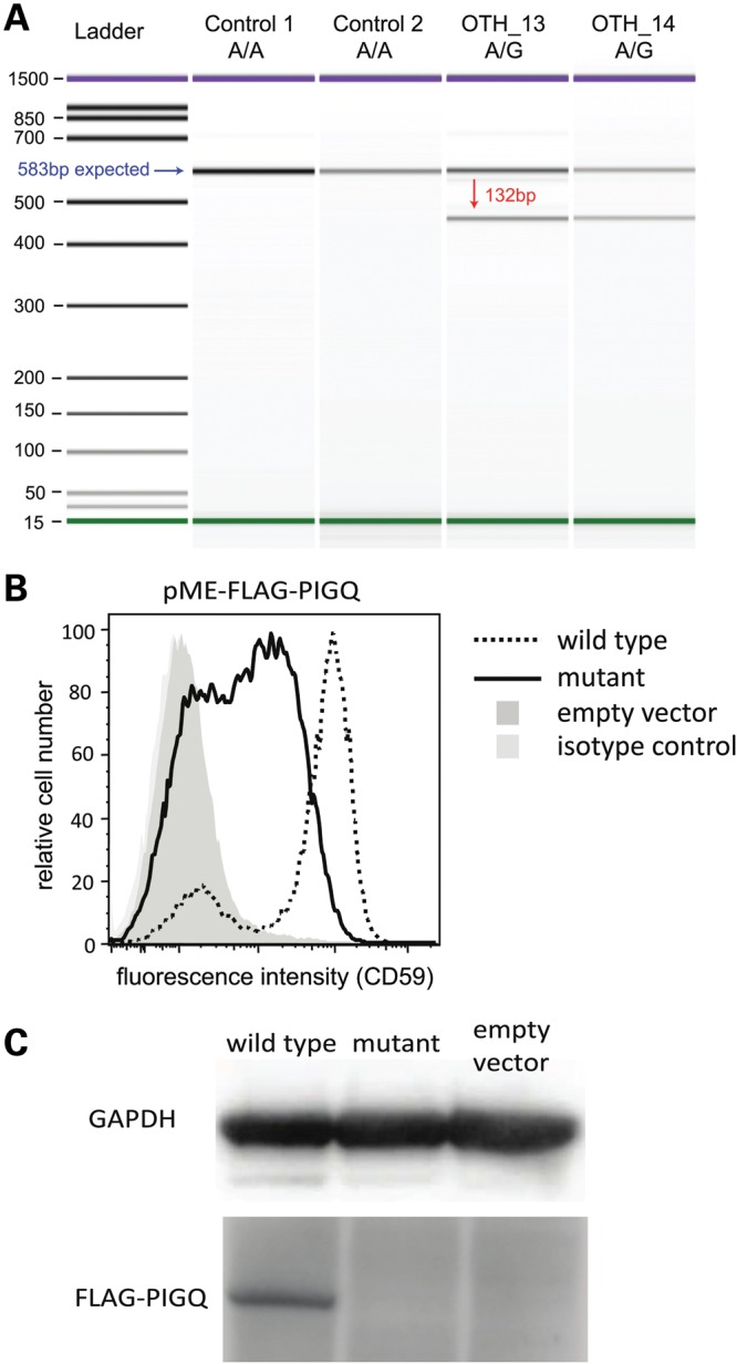
PIGQ splicing mutation in Patient 4. (A) The variant causes skipping of exon 3. This image shows the Bioanalyzer gel from an RT-PCR (see Materials and Methods) and demonstrates the presence of two PIGQ transcripts in the heterozygous parents (OTH_13, OTH_14). The blue arrow indicates the band expected from the annotated transcript, and the red arrow that expected from the skipping of exon 3. (B) Severely decreased functional activity of the mutant PIGQ. PIGQ-deficient CHO cells were transiently transfected with WT or mutant PIGQ cDNA (lacking exon 3). Restoration of the surface expression of CD59, a GPI-anchored protein, was assessed by flow cytometry after staining with anti-CD59 antibody. The mutant PIGQ did not restore the surface expression of CD59 as efficiently as the WT. X axis, fluorescence intensity corresponding to CD59 expression level per cell; Y axis, relative cell number. (C) The expression of mutant protein was greatly decreased and could not be detected by western blotting.