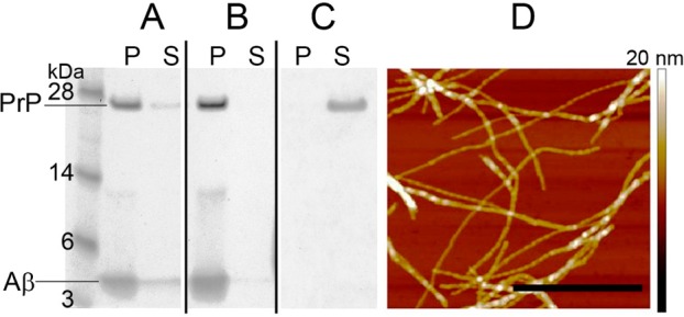
Figure 2.

Cosedimentation of PrP with Aβ1–42 fibrils. PrP (2 μM) was incubated in the absence or presence of intact Aβ1–42 fibrils or fibrils fragmented by sonication (50 μM in each case). The samples were subsequently centrifuged under the conditions allowing sedimentation of Aβ fibrils, and the pellets and supernatants were analyzed by SDS-PAGE. (A) PrP preincubated with intact Aβ1–42 fibrils; (B) PrP preincubated with fragmented Aβ1–42 fibrils; (C) PrP alone. Symbols P and S refer to pellet and supernatant, respectively. (D) AFM image of intact Aβ1–42 fibrils used in these experiments is shown to demonstrate that these preparations are indeed highly enriched in fibrillar aggregates. The scale bar represents 0.8 μm.
