Figure 3.
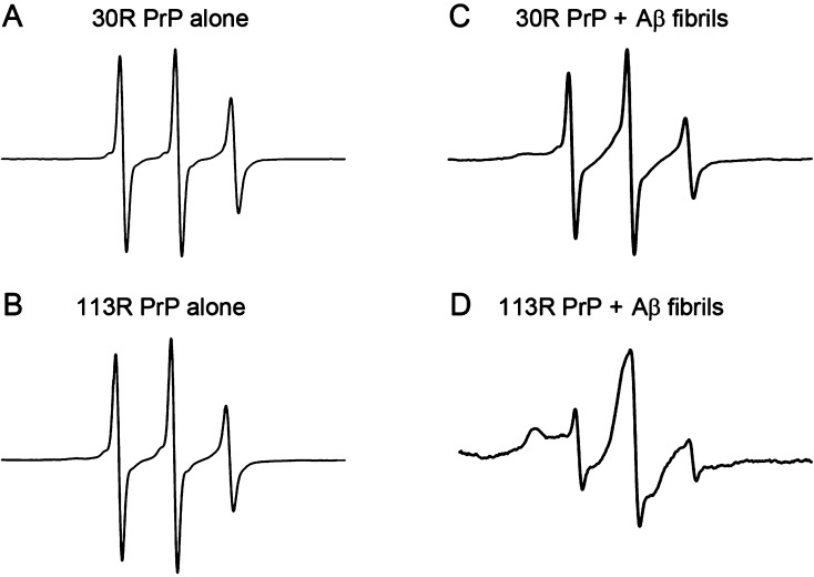
Binding of PrP to Aβ1–42 fibrils as probed by EPR spectroscopy. Representative EPR spectra for 30R PrP and 113R PrP (A and B) alone or in the presence of Aβ1–42 fibrils (C and D). The concentration of PrP and Aβ1–42 was 2 and 170 μM, respectively. Note that the EPR spectrum obtained for 113R PrP in the presence of Aβ1–42 fibrils (D) is characteristic of a highly immobilized spin label, whereas the immobilized component in the spectrum of 30R PrP incubated with the fibrils (low-field shoulder in panel C) is very weak.

