
Figure 6.
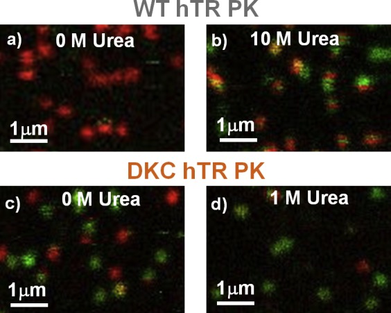
Images of surface-immobilized molecules for the wild-type (a, b) and dyskeratosis congenita (c, d) pseudoknots. Each diffraction-limited fluorescence spot represents the location of an individual dually labeled RNA molecule. The urea dependence of the images yields kinetic information about the two different pseudoknot constructs (see text for details).