Fig. 7.

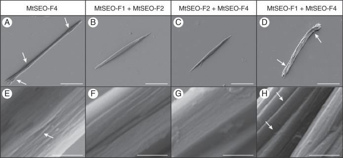
Scanning electron micrographs of artificial forisomes expressed in N. benthamiana leaves. (A) MtSEO-F4 protein bodies are thin in the centre and appear more rod-like than spindle-shaped, but single-fibril bundles can still be discerned at the tips (arrows). (B, C) MtSEO-F1 + MtSEO-F2 (B) and MtSEO-F2 + MtSEO-F4 (C) artificial forisomes possess the typical spindle shape. (D) MtSEO-F1 + MtSEO-F4 protein bodies comprise loosely attached fibres with distinct inter-fibre spaces (arrows). (E) Higher magnification of MtSEO-F4 reveals shallow cavities (arrow) in the protein body. (F, G) The surface of MtSEO-F1 + MtSEO-F2 (F) and MtSEO-F2 + MtSEO-F4 (G) artificial forisomes is tightly packed. (H) Magnification of an MtSEO-F1 + MtSEO-F4 protein body shows distinct fibres with large intervening cavities (arrows). Scale bars: (A–D) = 10 μm, (E–H) = 500 nm.
