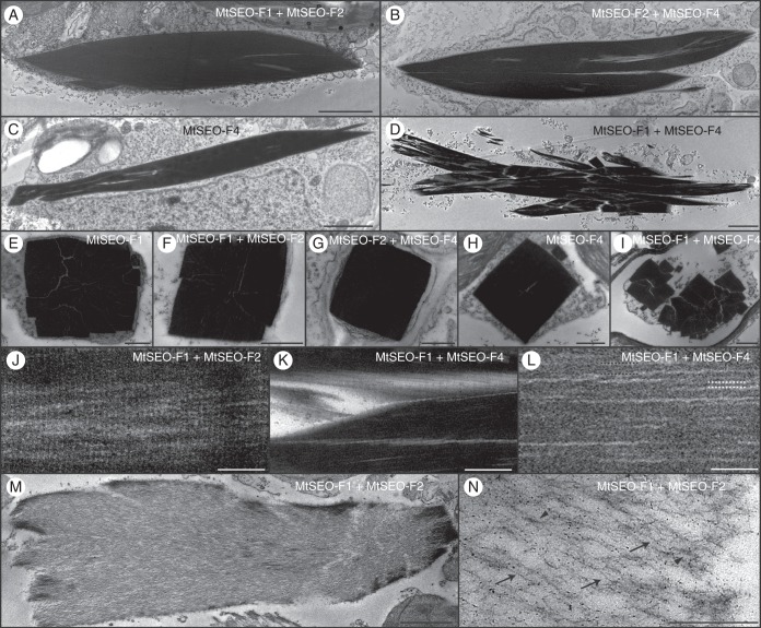
Fig. 8.
Representative transmission electron micrographs of artificial forisomes expressed in N. benthamiana leaves. (A–D) Longitudinal sections of condensed protein bodies following the combinatorial expression of MtSEO-F subunits. MtSEO-F1 + MtSEO-F2 (A) and MtSEO-F2 + MtSEO-F4 (B) protein bodies are densely packed, whereas MtSEO-F4 protein bodies (C) reveal some spaces. MtSEO-F1 + MtSEO-F4 protein bodies (D) reveal even more spaces between dense fibril bundles. (E–I) Cross-sections of condensed protein bodies following the expression of MtSEO-F1 (E), MtSEO-F1 + MtSEO-F2 (F), MtSEO-F2 + MtSEO-F4 (G), MtSEO-F4 (H) and MtSEO-F1 + MtSEO-F4 (I). With the exception of (I), all protein bodies were characterized by a cuboid outline and dense packing. In MtSEO-F1 + MtSEO-F4 protein bodies (I), the fibre bundles were separated from each other. (J) Higher magnification of a longitudinal section of the MtSEO-F1 + MtSEO-F2 protein body revealed a fine cross-striation pattern perpendicular to the forisome body. (K, L) Higher magnification of a longitudinal section of the MtSEO-F1 + MtSEO-F4 protein body. (K) Coarse cross-striation. (L) Fibrils (black dotted lines) and filaments (white dotted lines). (M, N) Expanded MtSEO-F1 + MtSEO-F2 protein body following the application of Ca2+ during embedding. (M) The outline of the protein body remains condensed, indicating that the expansion proceeds from the central body towards the periphery. (N) Single filaments (arrows) and fibrils (arrowheads) are distinguishable. Scale bars: (A–D, M) 2 μm, (E–I, K, N) 500 nm, (J, L) 100 nm.

