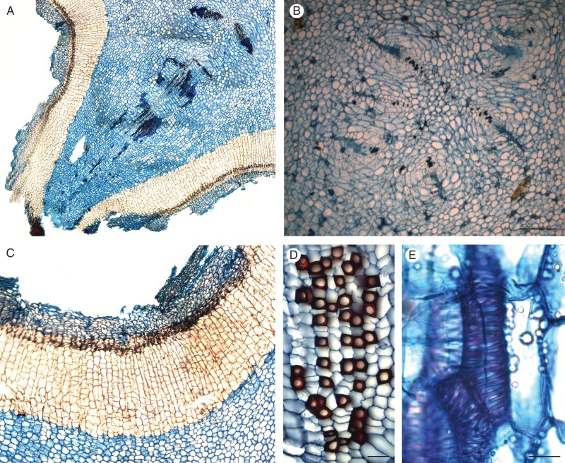
Fig. 5.
Details of anatomy of Hydnora. (A) Transverse section of an active bump of H. visseri, showing the developing vascular system of a new branch, originating from the vascular traces within the ridge; (B) transverse section of six vascular traces within a bump of H. visseri (scale bar = 500 μm); (C) transverse section of the periderm of H. visseri, including the primary cortical tissues; (D) transverse section of the xylem of H. longicollis, showing the lignified vessels (dark red) and the surrounding axial parenchyma (scale bar = 50 μm); and (E) tangential view of vessel elements of H. visseri (scale bar = 25 μm). Sections are stained with Astra blue-safranin.

