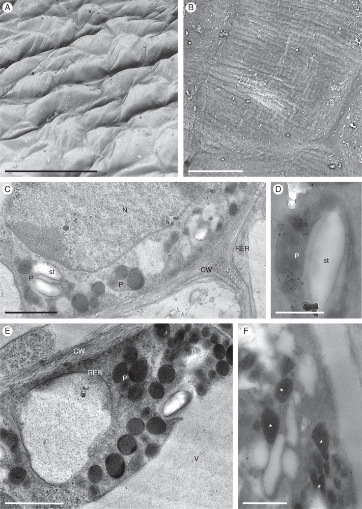
Fig. 12.
Cyrtochilum meirax: SEM and TEM. (A) Cuticular surface of epidermal cells of the callus lacking obvious secreted material. (B) Detail of the finely striate cuticle lacking blisters and cracks. (C) Highly granular and vesiculate parietal cytoplasm containing a large nucleus and plastids. (D) Plastid containing few internal membranes, as well as a starch grain and numerous plastoglobuli. (E) Large plastoglobuli may occur within plastids. (F) Osmiophilic bodies (asterisks), not unlike plastoglobuli, may also occur in the cytoplasm. Scale bars = 100 μm, 10 μm, 2 μm, 1 μm, 2 μm, 500 nm, respectively. CW, cell wall; N, nucleus; P, plastid; RER, rough endoplasmic reticulum; st, starch; V, vacuole.

