Fig. 5.
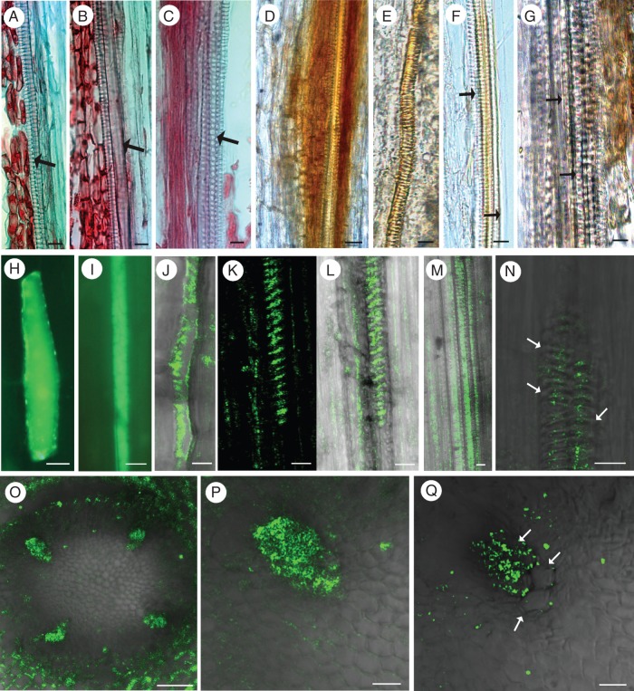
Pioneer root stele comparative anatomy of consecutive root segments (longitudinal sections stained with Safranin and Fast Green) showing all stages of the first tracheary elements in primary xylem development (A–C, arrows), and stages corresponding to those used for bioimaging of hydrogen peroxide (H2O2; D–G) and nitrogen oxide (NO; H–Q) during xylogenesis. Detection of H2O2 (DAB positive, represented by red-brown staining) in differentiating tracheary elements on the second day of root growth, in protoxylem vessels with annular or helical cell wall thickening (D, E) and in living cells directly adjacent to differentiating cells on the second day (D) or already differentiated vessels of 3-day-old roots (G). Note that H2O2 was not detected in large diameter functional vessels of stage 3 (F, G; arrows). Initial NO accumulation [represented by green fluorescence of the triazole molecule (DAF-2 T) formed from DAF-2DA] in single cells at the site of subsequent xylem formation on the first day of root growth (H–J), and NO appearance in the various stages of differentiating xylem on the second and third day of root growth (K–Q). Note the NO burst in non-lignified thin-walled cells with end walls visible – stage 1 (H–J) and the gradual disappearance of NO in the mature thick-walled xylem vessels – stage 3 (N, Q; arrows). Scale bars (A–N, P, Q) = 10 μm; (O) = 50 μm.

