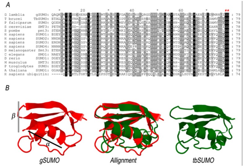
Figure 2.
Comparative analysis of SUMO proteins. (A) Multiple sequence alignment was constructed between the conserved domain of gSUMO and other 13 SUMO sequences, plus Human Ubiquitin. Dark shading show identical residues, while light shading shows similar residues between the respective sequences. Red asterisks indicate conserved GG motifs. (B) Structural alignment was constructed with alfa-helix (α) and beta-sheet (β) portions of the predicted 3D structure of gSUMO and the Chain A of the Protein Data Bank file 2K8H from T. brucei (TbSUMO).

