Figure 5.
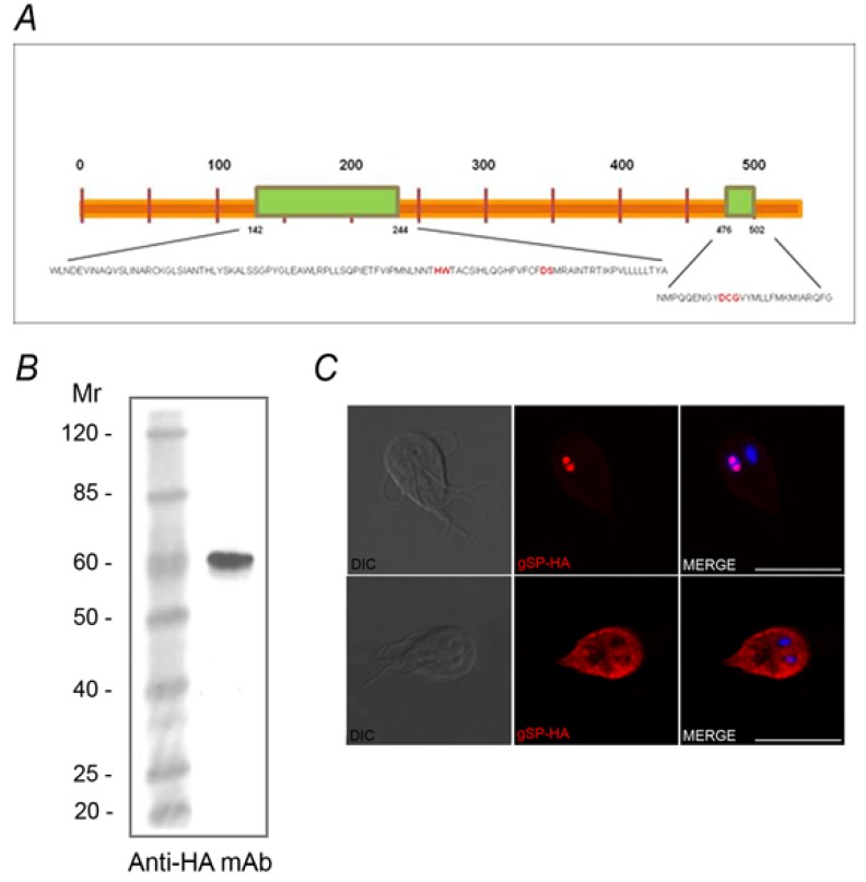
Giardia lamblia has one predicted gSP. (A) Schematic representation of the putative gSP containing two catalytic C-terminal domains (green) with the essential catalytic residues (red). (B) Western blotting showing a band of ~ 60 kDa (arrow) corresponding to free gSP in gSP-HA transgenic trophozoites (TT). (C) IFA using anti-HA mAb and confocal microscopy show gSP-HA (red) in the cytoplasm and nuclei (DAPI) of transfected Giardia trophozoites. WT: wild-type. Scale bar: 10 µm.

