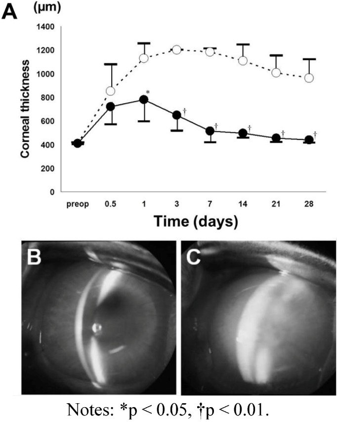
Figure 5.

Central corneal thickness (A) and anterior segment photographs (B,C) after transplantation of a DSAEK graft reconstructed with collagen and cultured HCECs [28]. The DSAEK graft using cultured HCECs and collagen (DSAEK group) or a bare collagen sheet (control group) are transplanted into rabbit corneas after stripping of the Descemet membrane. In the control group (open circles), the mean corneal thickness remains at around 1000 m for 28 days. In contrast, the mean corneal thickness gradually decreases in the DSAEK group (closed circles) and becomes significantly less than in the control group. There are significant differences of corneal thickness between the DSAEK and control groups on Days 1, 3, 7, 14, 21 and 28 using an unpaired t-test. (B) Representative anterior segment photographs obtained with a slit-lamp microscope at 28 days after surgery show a thin cornea without stromal edema in the DSAEK group; (C) while severe corneal edema is observed in the control group.
