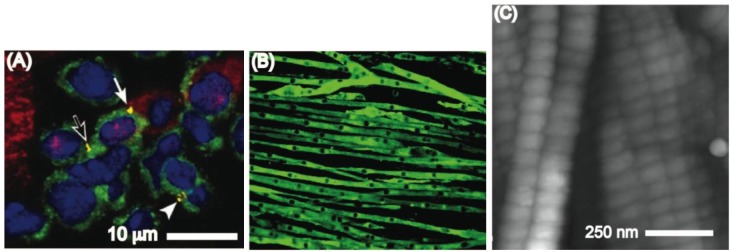
Figure 1.

(A) Cluster of cardiac stem cells (green) and lineage committed cells (red). The yellow regions stain for connexin 43 (Published with permission from PNAS [24]); (B) A fluorescent image in which myoblast actin filaments are stained green and the myoblasts nuclei are shown as dark elongated spots. Each myoblast tube is measured to be approximately 12.5 μm in diameter (Published with permission from Am Physiol Soc [25]); (C) Atomic force microscopy images of the D-band patterns on collagen I (Published with permission from Royal society Publishing [27]).
