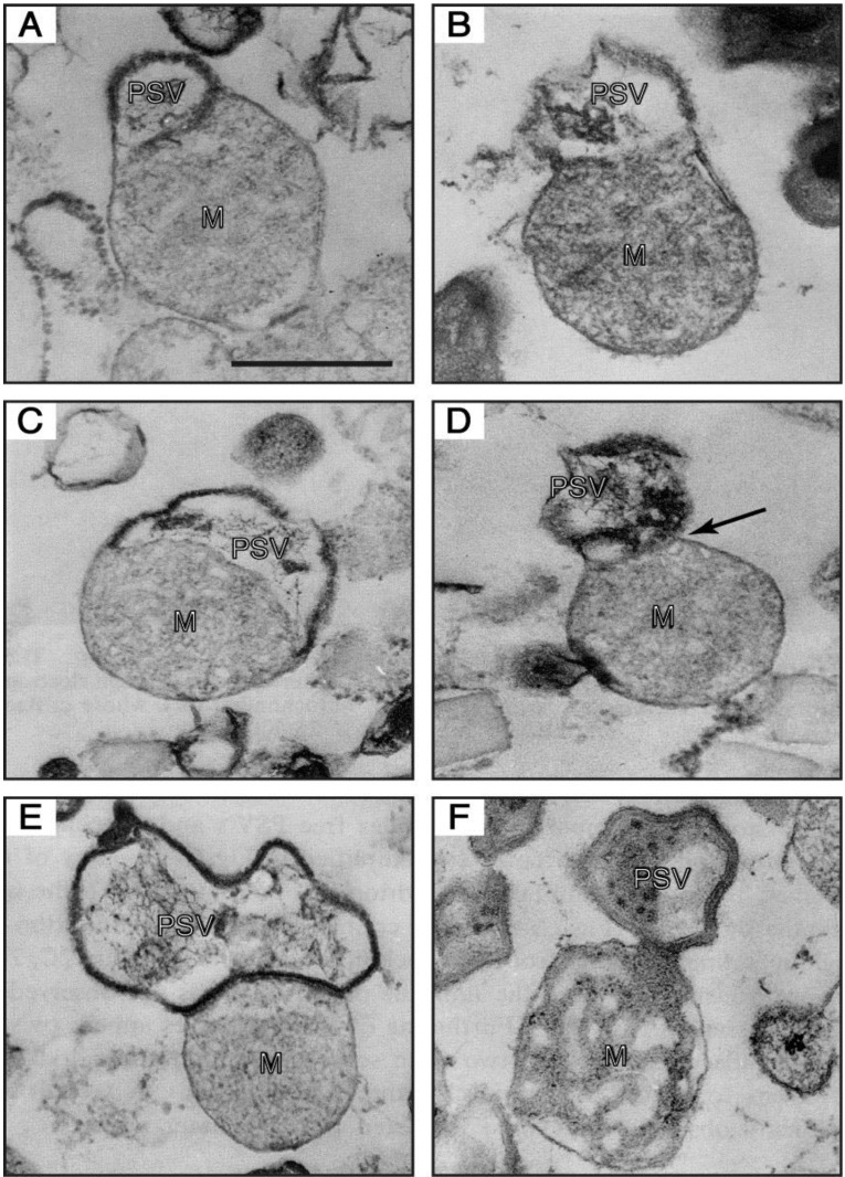
Figure 6.
Electron-micrographs of PSV-mitochondrion complexes present in a 47.5%–50% fraction (the intermediate fraction between a lighter pure mitochondria and a heavier pure PSV fractions) of a multilayered sucrose gradient for cell fractionation. (A,B) the outer membrane of a mitochondrion (M) is continuous with the unit membrane of a PSV. (C) a PSV and a mitochondrion are partitioned by a single membrane derived from either the PSV or the mitochondrion, and the electron-dense lining membrane of the PSV is not observed at the contact region. (D) a PSV appears twisted at the contact region (arrow). (E) a completely formed PSV is in close contact with a mitochondrion. (F) a mitochondrial part itself loses its inner structural integrity and seems to be transformed into the PSV. Bar, 500 nm. (From Maeda [49]).

