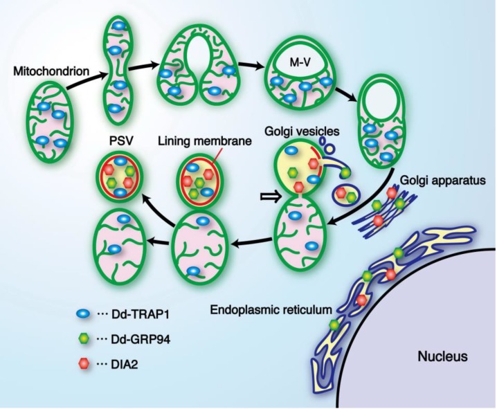
Figure 7.
A diagrammatic representation showing formation of the prespore-specific vacuole (PSV) from a mitochondrion and Golgi vesicles. Prior to PSV formation, mitochondria in differentiating prespore cells undergo drastic transformation to form a sort of vacuole (M vacuole), and some Dd-TRAP1 molecules translocate into the M vacuole. Subsequently, Golgi vesicles containing DIA2, Dd-GRP94 and other materials required for PSV formation fuse with the M vacuole, thus resulting in formation of the lining membrane (red) and the internal fibrous structure in the M vacuole. The mitochondrion-PSV complex is eventually twisted at the junction (arrow) and detached to form the respective organelles. Interestingly, almost all of the DIA2 molecules are selectively translocated to PSVs (possibly M vacuoles) in differentiating prespore cells, and seem to be required for exocytotic secretion of PSVs to form the outer-most membrane of spore cell wall [15]. M-V, M vacuole; GRP94, glucose-regulated protein 94 (endoplasmic reticulum Hsp90). (Slightly modified from Maeda [12]).

