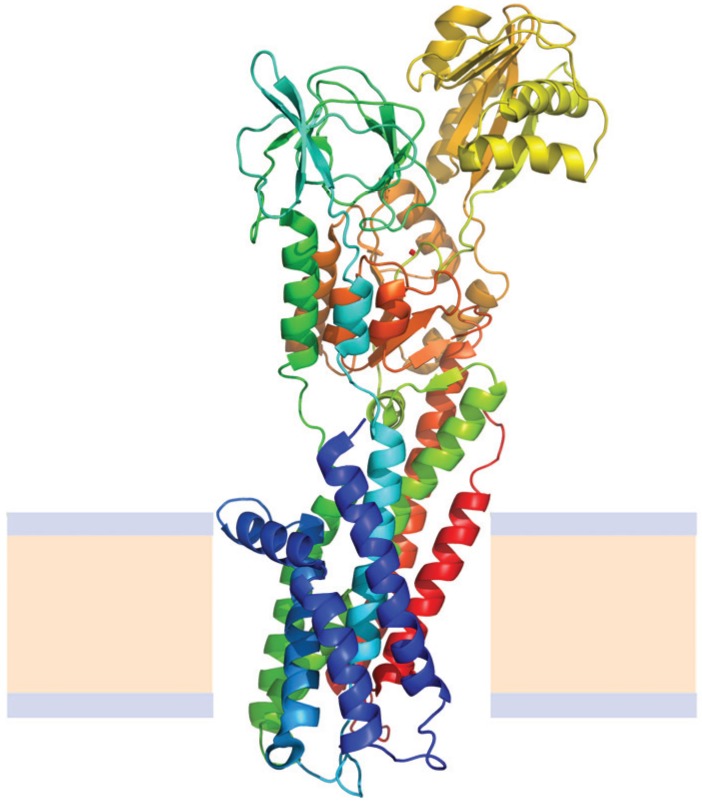
Figure 7.
Structure of the Cooper transport ATPase from Legionella pneumophila (Protein Data Bank ID 3RFU). Alpha helices are represented by coiled ribbons, beta sheets by arrows and protein loops are shown as thin lines. The beige area represents the hydrophobic core of the membrane, while the light-indigo regions refer to the hydrophilic head groups of phospholipids.

