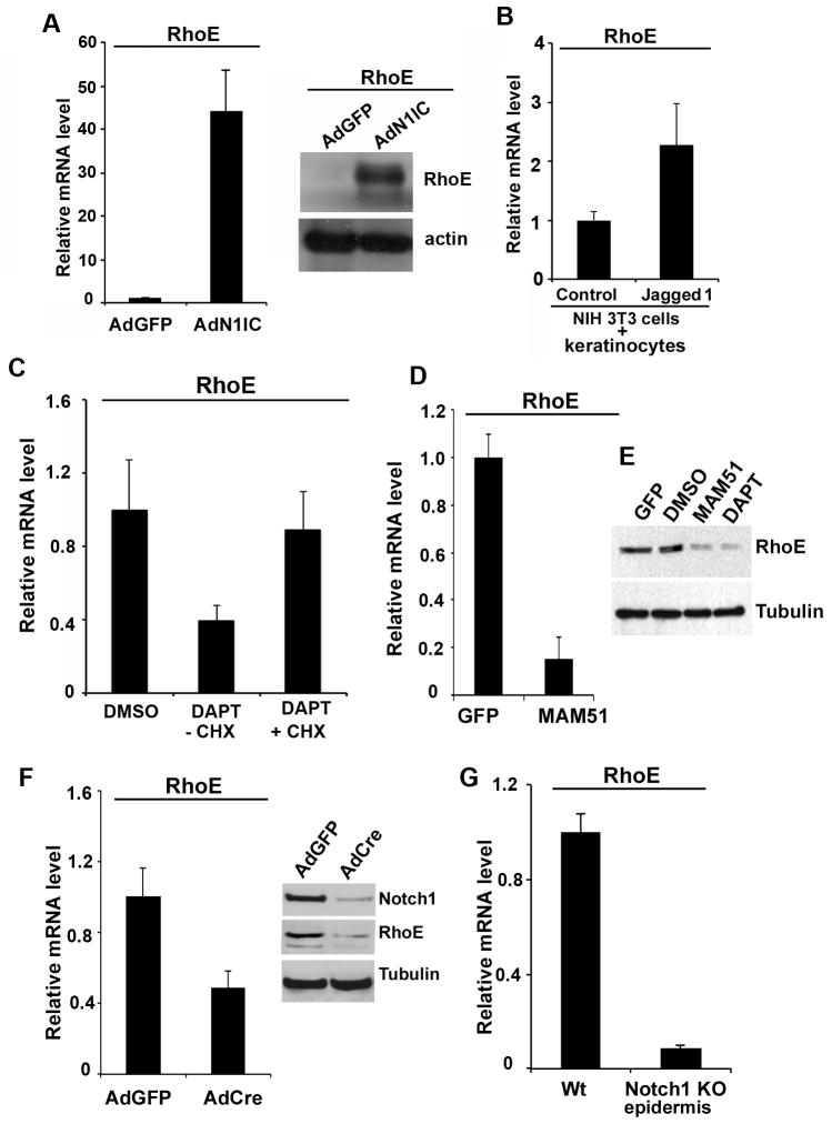
Figure 2. RhoE expression is regulated by Notch1 in keratinocytes.
A, Notch activation induces RhoE expression. HKC were infected with AdN1IC or control AdGFP, followed by assessment of RhoE mRNA levels by real time RT-PCR (left panel) and protein levels after Western blot. B, HKC were co-cultured with control mouse NIH 3T3 fibroblasts or NIH 3T3 fibroblasts stably expressing full-length Jagged1 for 48 hours, followed by real-time RT-PCR analysis of RhoE mRNA levels. C, HKC were treated with 10 μM DAPT or DMSO vehicle control for 24 hrs, after that DAPT was washed out with PBS and cell culture medium and cells were treated with cycloheximide for 2. RhoE mRNA levels were then assessed by real time RT-PCR analysis. D, MAM51-mediated inhibition of Notch transcription suppresses RhoE expression. (E) Western blot analysis of RhoE protein levels in MAM51 overexpressing or DAPT treated primary HKC with tubulin as a loading control. F, Primary mouse keratinocytes from homozygous Notchloxp/loxp were infected with AdCre or AdGFP control. At 72 hours after infection, cells were analyzed either by real time RT-PCR analysis and immunoblotting for RhoE expression levels using 36B4 mRNA levels or γ-tubulin protein levels for normalization, respectively. The deletion of the Notch1 gene was confirmed by immunoblotting with a Notch specific antibody. G, Real time RT-PCR analysis was used to analyze RhoE mRNA levels, using mouse GAPDH for internal normalization. Relative mRNA levels are presented as a fold change as compared to the control condition. Error bars represent Standard Deviations, calculated based on three independent measurements.

