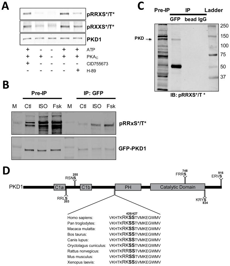
Figure 4. PKA-dependent phosphorylation of PKD.
(A) In vitro phosphorylation of purified PKD with the catalytic subunit of PKA. Phosphorylation is measured with 2 different PKA consensus motif antibodies (RRXS*/T* top and RXXS*/T* bottom). (B) ISO- and Forsk-induced phosphorylation of immunoprecipitated GFP-PKD1 measured with a PKA consensus motif antibody (RRXS*/T*). Blots are probed with GFP for loading control. (C) Control immunoprecipitations (beads only and control IgG) demonstrate specificity of the PKD pull-down. (D) Potential PKA phosphorylation sites on mouse PKD1 sequence.

