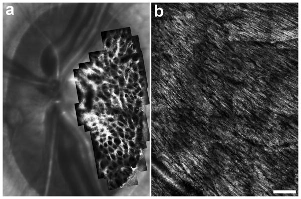
Figure 2.

Imaging the lamina cribrosa (a) and retinal nerve fiber layer (b). Image in (a) is a montage from a 5-year old normal rhesus macaque monkey, courtesy of Jason Porter, PhD and Kevin Ivers at the University of Houston College of Optometry. The image in (b) is a montage of the nerve fiber layer in a 62-year-old male with normal vision. Scale bar is 200 μm.
