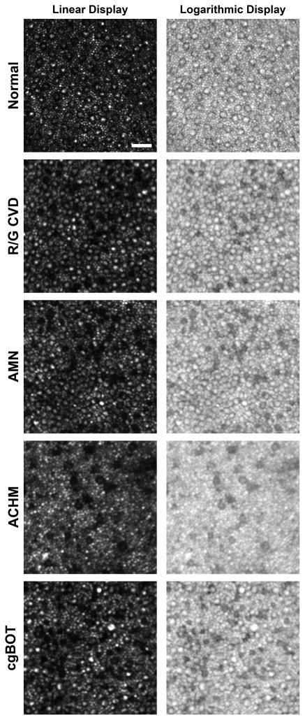
Figure 4.
Non-waveguiding cones in retinal disease. Shown are perifoveal images (linear and logarithmic display) of the photoreceptor mosaic for a subject with normal vision and 4 subjects with various retinal disorders. Normal cones at this eccentricity (~10 degrees from fixation) have a bright reflective center surrounded by a dark ring, where the extent of the dark area represents the inner segment diameter. Numerous cones devoid of the central reflective profile can be seen in a subject with red-green color blindness due to an LIAVA opsin mutation (R/G CVD),44 a subject with acute macular neuroretinopathy (AMN),45 a subject with achromatopsia (ACHM),43 and a subject with vision loss as a result of closed-globe blunt ocular trauma (cg-BOT). This altered reflectivity profile may indicate altered outer segment morphology. Scale bar is 20 μm.

