Figure 5.
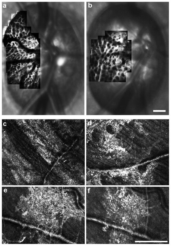
AOSLO imaging in glaucoma. Panel (a) shows an image of the anterior lamina cribrosa surface from the eye of a normal rhesus macaque prior to induction of experimental glaucoma, and panel (b) shows the laminar surface of the same eye at an early stage of experimental glaucoma showing an alteration to laminar beam and pore geometry. Images courtesy of Jason Porter, PhD and Kevin Ivers at the University of Houston College of Optometry. In human patients with glaucoma, drastic changes in reflectivity and striation in the peripapillary NFL corresponding to what would be clinically described as arcuate defects can be seen (c). Panels (d–f) illustrate hyper-reflective structures residing near the RNFL.61 These features were observed over areas of reduced visual sensitivity, and were not visible on SD-OCT or fundus photography. A four-month follow-up in one patient revealed significant changes in both the extent and appearance of the hyper-reflective patterns (e, f). Scale bars are 200 μm.

