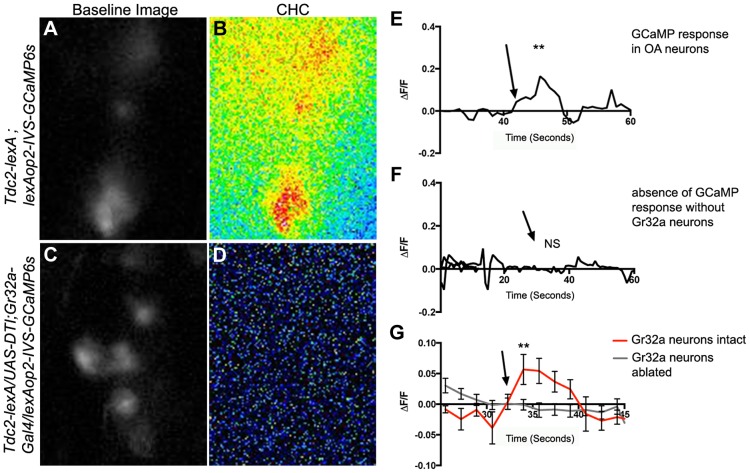
Figure 4. Male CHCs evoke intracellular Ca2+ responses in OA neurons that are dependent on Gr32a neurons.
(A) Greyscale image (background subtracted) of GCaMP3 fluorescence in OA neurons located within the SOG in a Tdc2-lexA;lexAop2-IVS-GCaMP6s male. (B) Pseudocolored subtraction image demonstrating an increase in fluorescence in response to male CHC application. (C) Greyscale image (background subtracted) of baseline fluorescence in the SOG of a male with Gr32a neurons eliminated (Tdc2-lexA; UAS-DTI;Gr32a-Gal4/LexAop2-IVS-GCaMP6s). (D) No changes in fluorescence are observed in the pseudocolored subtraction image of OA SOG neurons when male CHC extract is administered to the legs of males lacking Gr32a neurons. (E) A representative calcium signal trace of OA neurons expressing GCaMP6s in panels A–B in response to male CHC extract application (arrow), unpaired t-test **p<0.006. (F) A representative trace demonstrating the lack of calcium response in OA neurons after male CHC extract application (arrow) to the legs of males without Gr32a neurons (Tdc2-LexA;UAS-DTI;Gr32a-LexA/20XLexAop2-IVS-GCaMP6s). (G) The average calcium response of eight regions of interest from five Tdc2-lexA;lexAop2-IVS-GCaMP6s males (red line) before and after male CHC administration (arrow, unpaired t-test, **p<0.0001). The gray line is the average calcium response of ten regions of interest from five Tdc2-LexA;UAS-DTI;Gr32a-LexA/20XLexAop2-IVS-GCaMP6s males in response to male CHC administration. No significant change in response was observed (unpaired t-test, p<0.0788). Error bars denote s.e.m.

