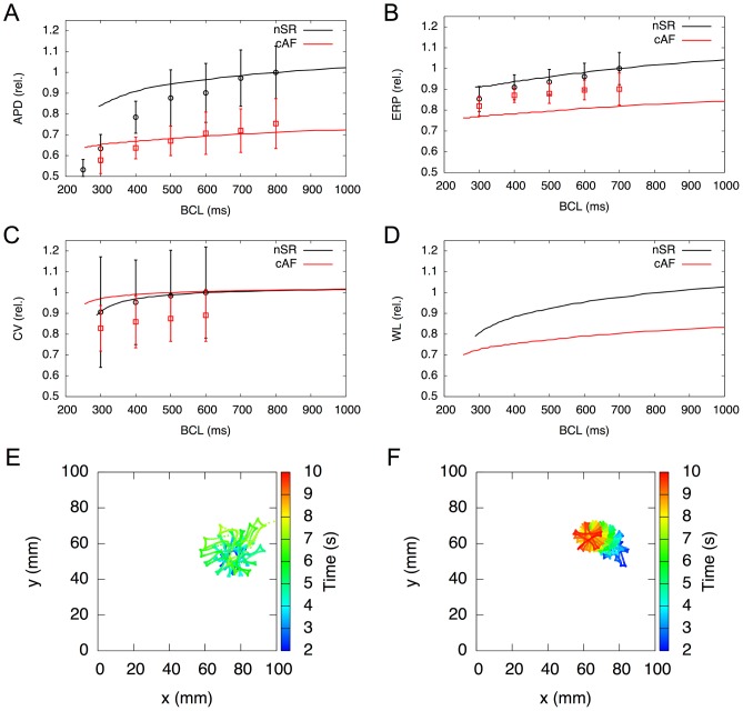
Figure 2. Electrophysiological properties in cAF tissue in silico.
A–D) Restitution properties in a 1D tissue beam, compared to in vivo results of Franz et al. [27] for action potential duration at 90% repolarisation (A), Yu et al. [28] for effective refractory period (B), Feld et al. [29] for conduction velocity (C) and wavelength (D). Simulation results are normalised to BCL = [0.8 0.7 0.6 1.0] s in (A–D), respectively. E&F) Mapping of rotor center trajectories after initiation in 2D tissue shows that in cAF (F) the meandering trajectory occupies a lot less space compared to nSR (E).

