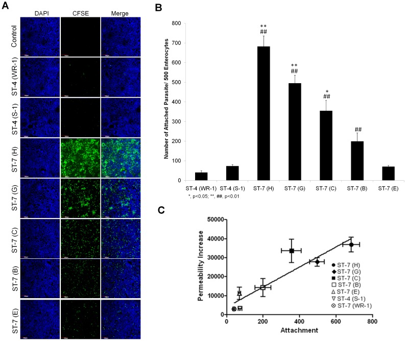
Figure 3. Blastocystis exhibits intra- and inter-subtype variation in attachment to Caco-2 cells.
(A) Representative confocal micrographs illustrating intra- and inter-subtype variation in Blastocystis attachment to Caco-2 cells. Caco-2 monolayers were grown to confluency on glass converslips and were then co-incubated with the same number of parasites of different strains of Blastocystis pre-stained with CFSE (green). Normal culture media was used as a negative control. After co-incubation, the non-attached parasites were washed away. The Caco-2 monolayers were then stained with DAPI and then were viewed using confocal microscope (Olympus Fluoview FV1000; Olympus, Japan). More green in the field represents more parasites attached to the monolayer. Both ST-4 strains adhere with a negligible number. An intra-subtype variation in the number of attachment within ST-7 is obvious. Isolates C, G, H appeared to attach at a much higher level than B and E to Caco-2 cells. Scale bar = 100 µm. (B) Graph representing number of Blastocystis parasites attached to host cells. ST-7 strains C, G and H exhibited a significantly higher number of attached parasites than ST-4 strains and ST-7 isolates B and E. *, p<0.05 vs. ST-7 (B, E); **, p<0.01 vs. ST-7 (B, E); ##, p<0.01 vs. ST-4. Each value represents a mean of six readings derived from 3 independent experiments. Error bar represents standard error. (C) Relationship between attachment and permeability increase by Blastocystis ST-4 and ST-7 parasites. The data points indicate individual strains. x and y error bars indicate the standard error for the respective measurements (n = 3). The R2 for the trend line shown is 0.8506, and the p value is 0.0031. There is a positive correlation between the level of attachment and permeability increase (R = 0.9223).

