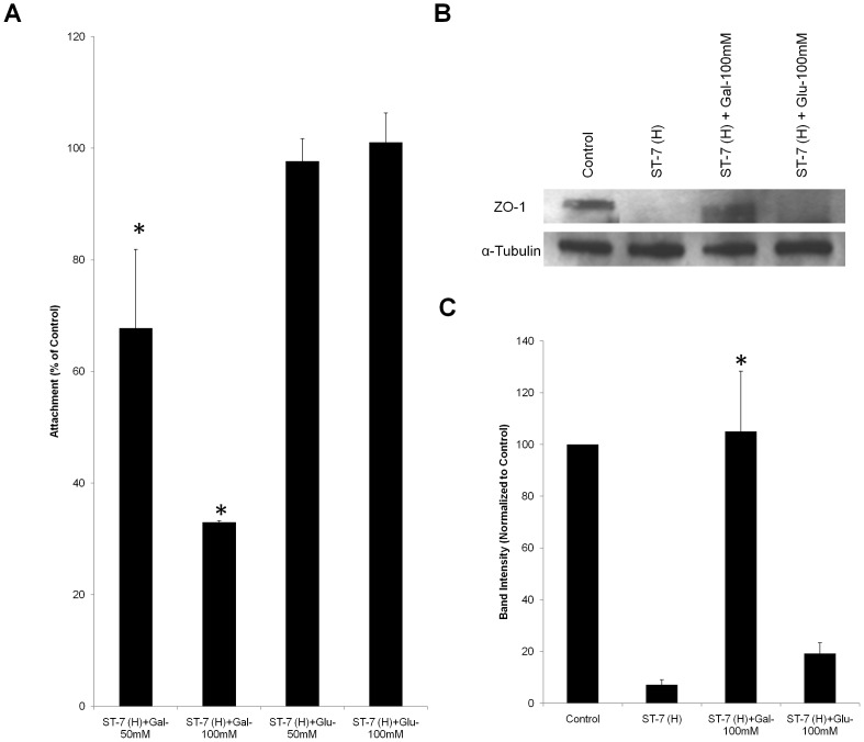
Figure 5. Inhibition of Blastocystis ST-7 (H) adhesion by galactose rescues Blastocystis ST-7 (H)–induced ZO-1 tight junction degradation.
(A) Dose-dependent inhibition of galactose on Blastocystis ST-7 (H) adhesion to Caco-2 monolayers. Blastocystis ST-7 (H) were incubated with epithelial cells in the presence of different concentrations of galactose and glucose (50 and 100 mM, respectively). A value of 100% was assigned to number of binding parasites without addition of sugars as control. The numbers of attached parasites with galactose addition were normalized to control.*, p<0.01 vs. control. (B) Representative western blot analysis of ZO-1 level in Caco-2 epithelium. Caco-2 monolayers were infected with Blastocystis ST-7 (H) in the presence of saccharides galactose and glucose at 100 mM and incubated for 1 h. Monolayers were washed and prepared for western blotting. Normal culture media with no sugar addition was used as the negative control. Galactose rescued Blastocystis–induced ZO-1 tight junction degradation. (C) Quantification of ZO-1 levels through densitometry analysis of Western blot radiographs. Densitometric values of ZO-1 signals were quantified and expressed as the ratio to α-Tubulin. ZO-1 degradation was significantly rescued by addition of galactose. *, p<0.01 vs. ST-7 (H)-treated sample. Values are the means ± standard errors from data of three experiments. Error bars represent the standard errors. Glu, glucose; Gal, Galactose.

