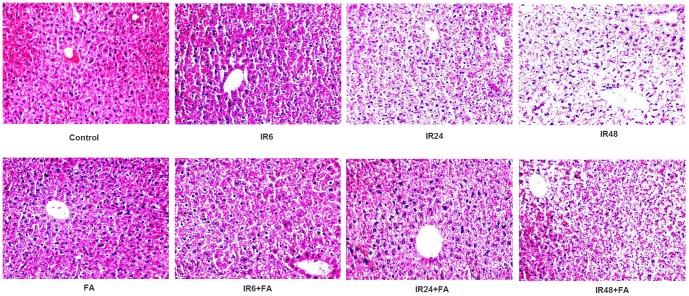
Figure 6. Photo micrograph from light microscope of mice liver sections. Sections were stained with haematoxylin and eosin.
Magnification, X 200: Control: without any treatment, IR6, IR24 and IR48: mice irradiated with 10 Gy γ -radiation, FA: mice treated with FA (50 mg/kg bodyweight) for 5 days and IR6+FA, IR24+FA and IR48+FA: FA treated plus irradiated.

