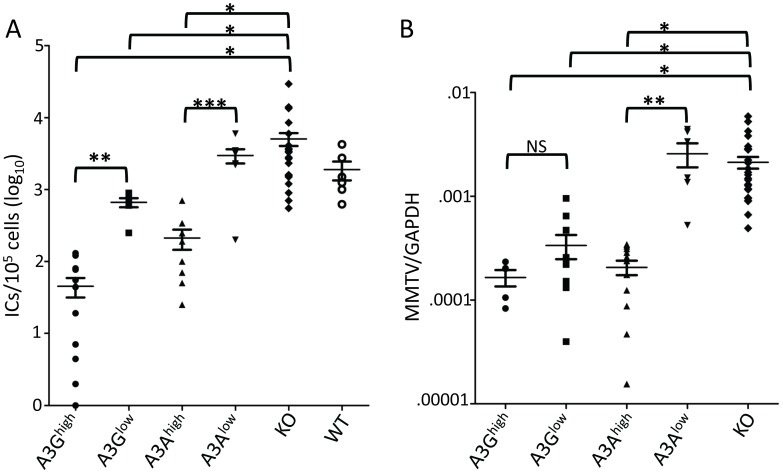
Figure 3. A3A and A3G restrict murine retrovirus infection in vivo.
A) Newborn mice were infected with M-MLV and 16 days post-infection, virus titers in spleens were measured. Each point represents the titer obtained from an individual mouse; the average for each group is shown by a horizontal bar. The transgenic mice were derived from 2–3 litters each; the knockout mice are the littermates of the transgenic mice. N = 12 A3Ghigh, 6 A3Glow, 10 A3Ahigh, 7 A3Alow, 42 KO and 6 WT mice. B) Five day old mice were infected with MMTV and 3 weeks post-infection, DNA was isolated from spleens and subjected to RT-qPCR with MMTV-specific primers. Each point represents the proviral DNA levels measured in splenic DNA from an individual mouse; the average for each group is shown by a horizontal bar. The transgenic mice were derived from 2–3 litters each; the knockout mice are the littermates of the transgenic mice. N = 5 A3Ghigh, 10 A3Glow, 12 A3Ahigh, 6 A3Alow and 24 KO mice. *, p≤0.0001, **, p≤.001, ***, p≤.01, NS, not significant (Mann-Whitney t test).

