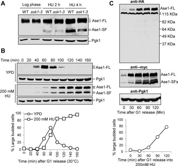
Figure 1. HU-induced expression of Ase1 short protein fragments.
(A) The expression of Ase1 in HU-treated WT and ask1-3 mutant cells. ASE1-13myc and ask1-3 ASE1-13myc cells were grown to log phase at 30°C and released to YPD containing 200 mM HU. Cells were collected at the indicated time points and protein samples were prepared for western blotting. Full length Ase1 migrates around 140 kDa and the short isoforms migrate around 105 kDa. Pgk1 protein migrates at 45 kDa and levels are shown as a loading control. (B) Expression of Ase1 isoforms in synchronized cells treated with HU. G1-arrested ASE1-13myc cells were released into YPD or YPD containing 200 mM HU. α-factor was restored in untreated cells to block the second round of cell cycle. Cells were collected at the indicated time points to detect the expression of Ase1 protein using western blotting. Budding index was used to indicate cell cycle stage and Pgk1 protein levels are shown as a loading control. (C) Ase1 short isoforms are not protein cleavage products. G1-arrested ase1Δ cells containing 3HA-ASE1-13myc plasmid were released into fresh selective media containing 200 mM HU and protein samples were prepared at the indicated time points. Western blotting with anti-HA and anti-myc antibodies was used to determine the expression of full length (Ase1-FL) and short isoforms (Ase1-SF) of Ase1 protein. Budding index was used to indicate cell cycle stage and Pgk1 protein levels are shown as a loading control. Asterisk indicates a non-specific band. Sell also Figure S1.

