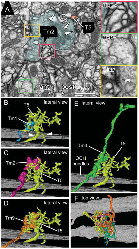
Figure 2.
Tm cell synaptic inputs to T5 dendrites. (A) EM of lobula stratum Lo1 showing a terminal of Tm2 surrounded by T5 dendrites expressing HRP over their membranes. Insets: HRP + (red) and HRP − (green) profiles; with presynaptic T-bar ribbon visible in Tm2 (yellow) providing input to HRP labeled T5 dendrites. (B–F) Representative pairs of Tm terminals overlapping the dendritic arbor of cell T5-1. Confirming its identity the latter has a bifurcated axon with the more slender branch going to the lobula plate, and its stouter partner connecting to the cell body (arrowheads in B). With T5-1 are lateral views of: (B) Tm1 (same terminal as Figure 1I); (C) Tm2 (same terminal as Figure 1J); (D) Tm9 (same terminal as Figure 1K); and (E) Tm4 (same terminal as Figure 1H). Background: bundles of horizontally directed chiasmal axons. (F) In a tangential plane, the overlap between neighboring T5 cells: T5-02 (orange), T5-01 (yellow) and T5-11 (green) with their color-coded synaptic inputs from four types of Tm input cells, Tm1, Tm2, Tm4 and Tm9 (between 1 and 3 cells per type, color coded as in Figure 3A–L).

