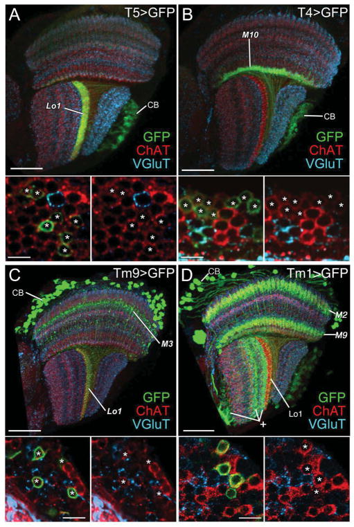
Figure 4.
T4, T5, Tm9, and Tm1 neurons express immunoreactivity to choline acetyltransferase (ChAT, red) but not vesicular glutamate transporter (VGlut, cyan). (A–D) T5, T4, Tm9, and Tm1 neurons are labeled with the mCD8::GFP membrane marker (green) using T5-Gal4, T4-Gal4, Tm9-Gal4, and Tm1-Gal4 drivers, respectively. (A) T5-GFP expression highlights a band of dendrites in lobula stratum Lo1 and cell bodies (CB) in the lobula plate cortex and their axons in the internal chiasma between lobula and lobula plate. (B) T4-GFP expression highlights a band of dendrites concentrated in medulla stratum M10 and cell bodies (CB) in the lobula plate cortex and their axons penetrating the lobula plate and entering the internal chiasma between medulla and lobula plate. (C) Tm9-GFP expression reveals somata (CB) in the medulla cortex, axons that penetrate the medulla neuropil, an arborization in stratum M3, and a band of terminals in lobula stratum Lo1. (D) Tm1-GFP expression reveals somata (CB) in the medulla cortex, dendrites that arborize in medulla strata M2 and M9, and axons that terminate in lobula stratum Lo1. The Gal4 line also labels uncharacterized neurons in the lobula (plus sign) and lobula plate. Lower panels: high-magnification views of the corresponding cell bodies (asterisks) in the cortex of the lobula plate (A, B) and medulla (C, D), distributed amongst cell bodies of other neurons (no asterisk). The left hand image of each pair shows all three labels; for clarity the green channel is omitted in the right hand panel, to show that all GFP expressing somata are also ChAT-positive. Scale bars: 30 μm in A–D; and 5 μm in lower panels.

