Figure 4.
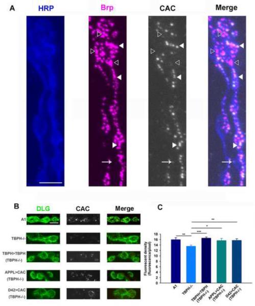
Localization and quantification of cacophony at the NMJ. A. Cacophony co-localizes with a pre-synaptic active site marker. Triple labeled image of the NMJ of muscle 6 showing motor neuron axons and terminals labeled with HRP (blue), the pre-synaptic active site marker bruchpilot (Brp, magenta) and cacophony (CAC, white). The fourth panel (Merge) is a merged image showing that cacophony is frequently co-localized with bruchpilot (closed arrow head) although occasionally we observed cacophony in the absence of bruchpilot (arrow) and bruchpilot in the absence of cacophony (open arrow head). B & C. Quantification of cacophony within the pre-synaptic terminals of motor neurons. B. Representative images of 1B terminals from larvae of the same genotypes shown in figures 2 and 3. The terminals were defined by staining with discs large (DLG, green). C. The cacophony fluorescence intensity within terminals was measured and showed a significant reduction in TBPH null mutants compared to the other genotypes (ANOVA, * p<0.05, ** p<0.01, *** p<0.001, n=3 animals each with at least 6 terminals). Scale bar is 5 μm in A and B.

