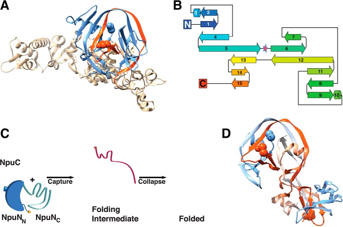
FIGURE 1.
Intein fold. A, T. kodakaraensis Pol-2 intein (Protein Data Bank (PDB) 2CW7) (9). The N-intein (blue) and C-intein (red) are shown. B, secondary structure topology map of Drosophila melanogaster Hog domain, a relative of inteins (PDB 1AT0) (6). The pseudo two-fold symmetry axis is highlighted with a star. C, capture and collapse folding mechanism of the Npu DnaE intein (12). The N-intein is blue (dark blue for NpuNN and light blue for NpuNC), the C-intein is red, and the extein-bearing termini are marked by orange circles. D, three-dimensional domain swapped dimer of a contiguous Npu DnaE intein variant (3).

