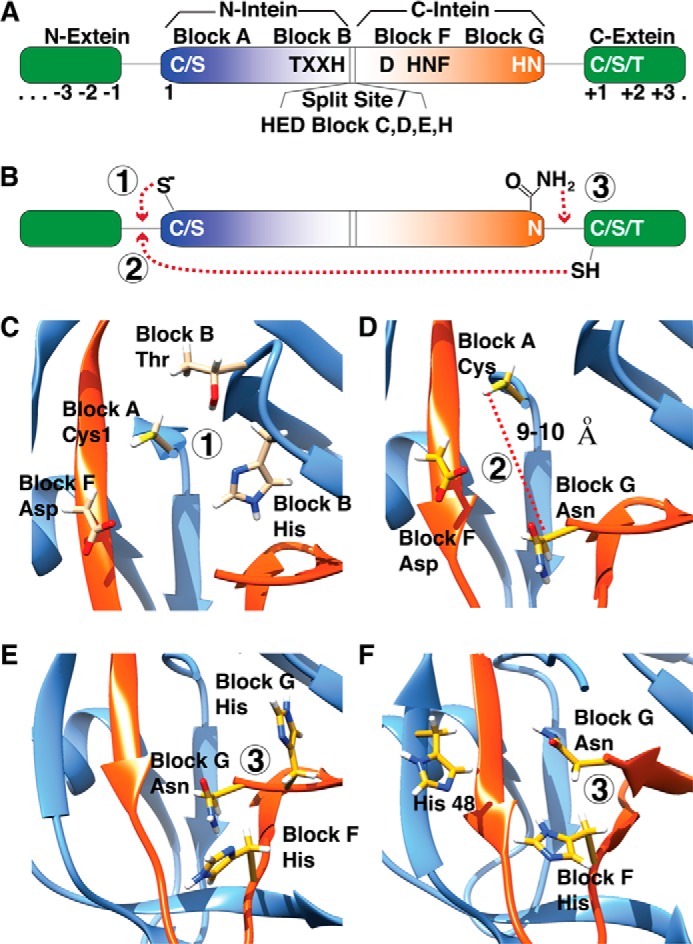
FIGURE 2.

Canonical splicing motifs and mechanism. A, intein is shown as a color gradient from blue to red, and exteins are show in green. The canonical motifs and corresponding blocks are shown. B, a simplified schematic of splicing reaction is shown; the splicing steps 1–3 are shown with circles. C–E, key residues, which take part in each splicing step, steps 1–3 in panel B, are highlighted as yellow sticks on the Mtu RecA intein (PDB 2L8L) (19). F, an alternate mechanism seen in DnaE inteins, lacking penultimate histidine, is shown on the Npu DnaE intein (PDB 2KEQ) (10).
