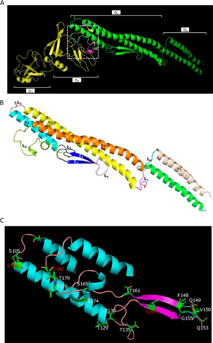
FIGURE 4.
Structural superposition of the flagellin FlaA (blue) and FlaB (purple) models with StFliC (yellow). The structural alignment and figure were produced with PyMOL. A, the domains were named according to StFliC. The sequences between two labeled residues (in FlaA and FlaB) represent the distinct portions among these proteins. B, the S. oneidensis FlaB domain structure according to StFliC with modifications. The structural alignment and figure were produced with PyMOL. C, the location of residues replaced by an alanine that caused a significant impact on motility. Residues in red represent the glycosylation sites.

