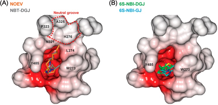
FIGURE 5.

Comparison of the binding of the PC compounds. Electrostatic potential surface of the surrounding of the active site pocket. A, NOEV (orange) complex structure is superimposed on NBT-DGJ (gray) complex. B, 6S-NBI-DGJ (green) and 6S-NBI-GJ (cyan) are drawn in a similar manner as in A.
