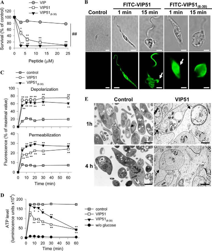
FIGURE 7.
The VIP derivatives induce the formation of disrupting membrane pores in L. major promastigotes. A, cell viability of L. major Friedlin clone V1 incubated with medium (control) or with various concentrations of VIP and the VIP derivatives determined by reduction of Alamar Blue and expressed as a percentage of untreated control samples. B, binding of the FITC-labeled VIP derivatives to L. major promastigotes (bottom panels). Top panels, Normarski images. Arrows point to sites of plasma membrane damage. Bars, 4 μm. C, variation in plasma membrane potential (top) and integrity (bottom) of parasites cultured with medium (control), VIP51, or VIP51(6–30). Data are expressed as a percentage of fluorescence intensity relative to that achieved by maximal depolarization or permeabilization. D, changes in ATP levels (expressed as luminescence units) in L. major promastigotes cultured with medium (control), VIP51, or VIP51(6–30). Parasites incubated in the absence of glucose were used as negative controls. E, ultrastructural analysis of the cell damage caused by VIP51 in L. major. The arrows point to membrane breakages (at 1 h) and depletion of electron-dense cytoplasmic material, severe cellular damage, and apparent leakage of the cytoplasmic content (at 4 h). Bars, 1 μm. The mean ± S.E. (error bars) (n = 3, each performed in duplicate) is shown. *, p < 0.05; **, p < 0.001 versus untreated parasites; ##, p < 0.001 versus VIP-treated parasites.

