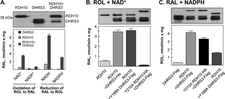
FIGURE 4.

Co-expression of WT or mutant DHRS3 and RDH10 in Sf9 cells. A, DHRS3 + RDH10 microsomes. B, RDH10 + Y188A DHRS3 or Y210RDH10 + DHRS3 microsomes incubated with 2 μm retinol (ROL) and 1 mm NAD+. C, RDH10 + Y188A DHRS3 or Y210RDH10 + DHRS3 microsomes incubated with 2 μm retinaldehyde (RAL) and NADPH. A-C (top panels), Western blot analyses of Sf9 microsomes (3–5 μg). In Sf9 cells DHRS3 protein appears as three bands when probed with either ProteinTech antibodies or our custom-made DHRS3 antibodies. HA-tagged Y210A RDH10-HA runs higher than RDH10. The His6-tagged DHRS3 and FLAG-tagged DHRS3 have similar molecular weight. B and C, bottom panels, reaction rates for microsomes (2 μg each). The results are representative of several measurements using the same preparation of microsomes. Note that both the oxidative and reductive activities are significantly higher for microsomes containing RDH10 + DHRS3. The results demonstrate that DHRS3 itself does not utilize NAD+ as a cofactor but its WT and mutant forms both activate the NAD+-dependent retinol dehydrogenase activity of RDH10, whereas WT and mutant RDH10 both activate the retinaldehyde reductase activity of DHRS3.
