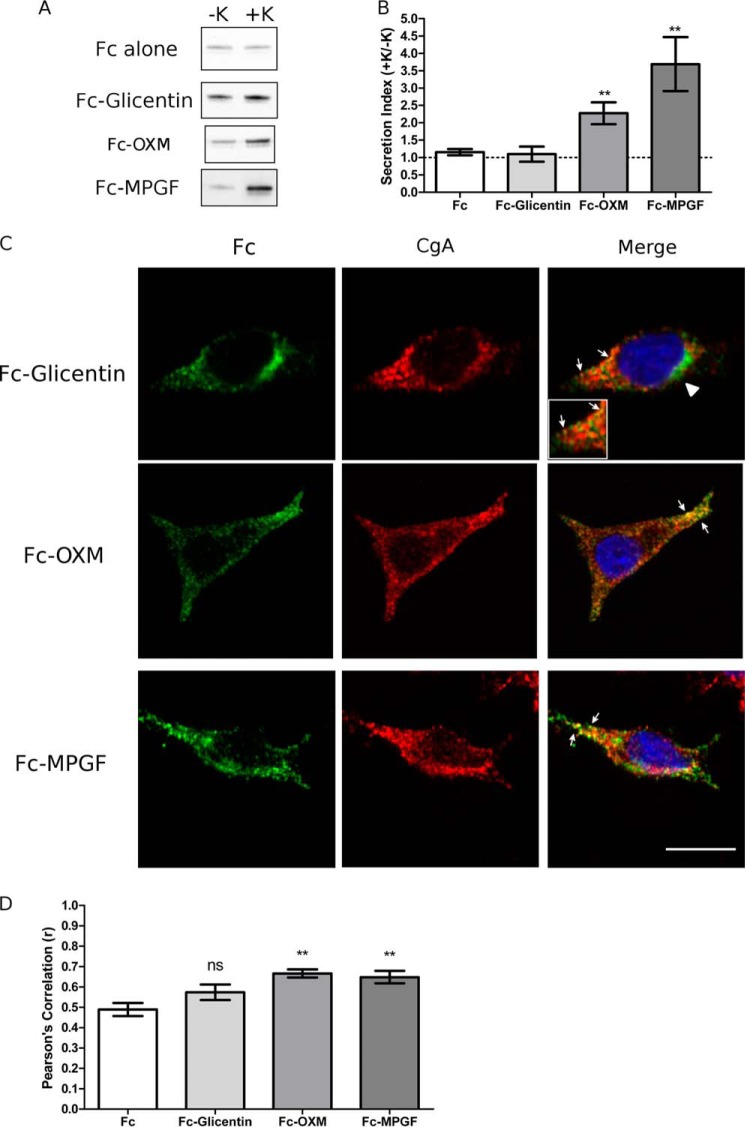
FIGURE 5.
MPGF is efficiently sorted into secretory granules by the sorting signal contained within GLP-1. PC12 cells were transfected with Fc-glicentin, Fc-MPGF, or Fc-OXM fusion constructs. A, Western blot analysis of regulated secretion. PC12 cells were incubated for 15 min without (−K; constitutive) and then with (+K; stimulated) 55 mm K+. Media were immunoprecipitated for Fc prior to Western blot analysis. Representative blots are shown (n = 6). B, secretion indexes from A. The dashed line indicates a secretion index of unity. Values are means ± S.E. (n = 6). **, p < 0.01 versus Fc alone. C, subcellular localization of Fc-glicentin, Fc-MPGF, and Fc-OXM fusion proteins. Arrows indicate co-localization of Fc with CgA. Arrowheads denote Golgi localization. Scale bar, 10 μm. D, PCC for co-localization between Fc and CgA. Values are means ± S.E. (n = 30–35). **, p < 0.01 versus Fc alone; ns, compared with Fc alone.

