
Figure 2. In vitro characterization of caspase-3/7-sensitive nano-aggregation fluorescent probe (C-SNAF).
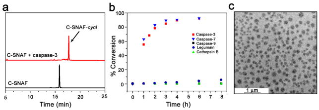
a, HPLC traces of C-SNAF in water (black, TR = 15.8 min) and the incubation of C-SNAF (25 μM) with recombinant human caspase-3 (4.9 × 10⁻³ U/ml) for 24 h at 37 °C in the caspase-3 buffer (red, TR = 17.7 min). b, The enzymatic reaction kinetics and specificity studies by longitudinal monitoring of % conversion of C-SNAF (25 μM) to C-SNAF-cycl after incubation with equal masses (0.735 μg/ml) of recombinant human caspase-3, caspase-7, caspase-9, cathepsin B, or legumain. c, TEM image of nano-aggregates after incubation C-SNAF (50 μM) with recombinant human caspase-3 (4.9 × 10⁻³ U/mL) overnight at 37 °C in caspase-3 buffer; scale bar, 1 μm.