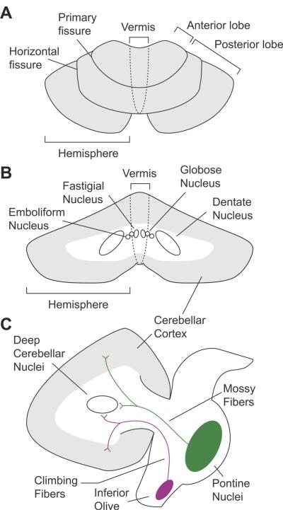
Figure 1.
Cerebellar anatomy. A) Top-down axial view. B) Coronal view of the deep cerebellar nuclei. C) Sagittal view highlighting the major afferent pathways into the cerebellum. Mossy fibers project to granule cells in the cerebellar cortex, and send collaterals to the deep cerebellar nuclei. Climbing fibers extend to Purkinje cells, and also have collateral projections to deep cerebellar nuclei. The axons of the deep cerebellar nuclei form the primary output channels away from the cerebellum, and to the brainstem and cerebral cortex.

