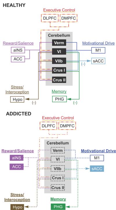
Figure 4. A cerebellar model for addiction.
The top panel highlights the cerebellum's role as a modulator of affective and cognitive function in the healthy brain. This schematic highlights the four major brain networks proposed to be affected by addiction (adapted from Volkow et al., 2010). The components within each network are color-coded to match the cerebral resting state networks in Figure 2 (adapted from Buckner et al., 2011). Arrows show the proposed direction of cerebellar down-modulation of specific brain regions within each circuit (Moulton et al., 2011). The bottom panel proposes how cerebellar dysfunction may impact the addicted brain. The black dashed boxes highlight structural degradation of specifically localized cerebellar structures with addiction. The dashed lines show disrupted functional connectivity and cerebellar modulation of specific brain regions related to executive control, reward/salience, motivational drive, and memory. Filled colored boxes and bold type represent brain circuits released from cerebellar modulation, leading to an uninhibited and sensitized neurological state. Note that all cerebellar outputs project through the deep cerebellar nuclei (refer to text for details). ACC, anterior cingulate cortex; aINS, anterior insula; DLPFC, dorsolateral prefrontal cortex; DMPFC, dorsomedial prefrontal cortex; Hypo, hypothalamus; M1, primary motor cortex; PHG, parahippocampal gyrus; sACC, subgenual anterior cingulate cortex; Verm, vermis; VI, cerebellar hemispheric lobule VI; VIIb, cerebellar hemispheric lobule VIIb.

