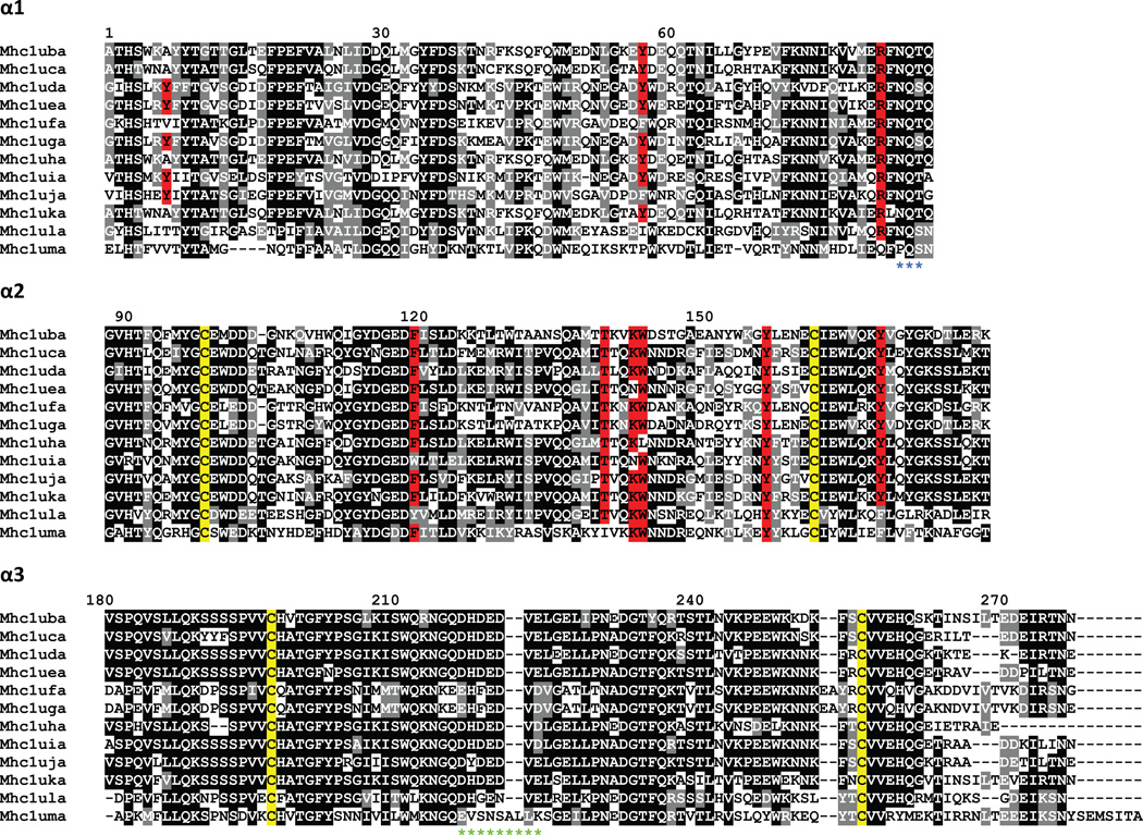
Figure 5. Alignment of MHC class I U lineage proteins.
The proteins encoded by the U lineage genes were organized into domains and aligned. Numbering of amino acids is based on the Mhc1uba sequence starting with the first residue of α1. Positions that are at least 50% identical are shaded in black and similar residues are shaded in gray. Conserved cysteine residues likely involved in the Ig-fold are highlighted in yellow. Residues implicated in peptide binding are highlighted in red. A potential N-linked glycosylation site at the end of α1 is indicated by blue asterisks below the alignment. A stretch of acidic residues in α3 that may associate with CD8 is indicated by green asterisks below the alignment.

