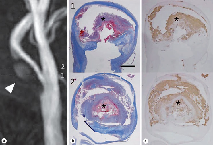
Fig. 2.
a Example of an HIS-positive plaque on MIP images of TOF MRA. Images were obtained from a 61-year-old man who had asymptomatic severe carotid artery stenosis. HIS in the plaque was observed on MIP images on TOF MRA (arrowhead). Numbers mean the slice level of MRA. Numbers in MRA and histology are matched as the same slice level. b The matching histology Masson's trichrome staining showed large regions of IPH (asterisk). c The antibody to glycophorin A also produced a strong signal in areas of hemorrhage (asterisk). Scale bar = 2 mm. Reprinted with permission from Yoshimura et al. [7].

