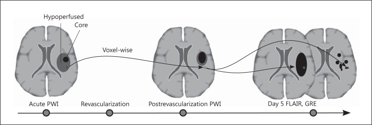
Fig. 2.
Schematic drawing of method for assessment of reperfusion injury in stroke patients. PWI = Perfusion-weighted images. Perfusion-weighted MRI images are acquired during the acute phase and after revascularization attempts. Reperfusion injury assessed by voxel-based comparison of the reperfused area and then subsequently undergoing irreversible cell death as measured by FLAIR sequence and/or hemorrhage as demonstrated by GRE sequence.

