Fig. 4.
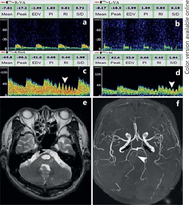
Reversed BA flow with proximal basilar occlusion. a, b Low MFV and high-resistance flow in the atlas segment of both VAs detected with continuous-wave doppler sonography at the mastoid is consistent with severe distal steno-occlusive disease. c Flow spectra obtained from the right PCoA is away from probe and with transmitted tapping (white arrowhead) of the right common carotid artery in the neck, indicating provide blood supply for posterior circulation. d Reversed flow is observed in BA with transmitted signals during tapping of the common carotid arteries in the neck (white arrowhead), indicating supply by carotid system. e Axial T2-weighted MRI shows brainstem infarction. Flow void of BA disappeared and filled with high signal (black arrowhead), indicating occlusion of the BA. f MRA shows prominent PCoA (black arrows) and proximal occlusion of BA (white arrowhead).

