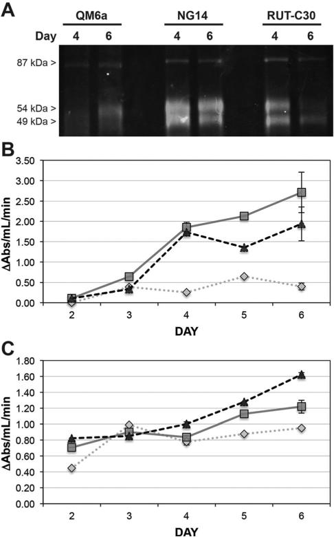
Fig. 3.
Comparison of cellulase and xylanase enzyme activity by GH-ABP labelling and Azo-CMC and AWAX assays. (A) Fluorescent gel analysis of GH-ABP (75 μM) labelling of days 4 and 6 culture of T. reesei strains QM6a, NG14, and RUT-C30. (B) Determination of cellulase activity in each T. reesei strain by the Azo-CMC assay. (C) Determination of xylanase activity in each T. reesei strain by the AWAX assay. For both assays measurements on days 2, 3, and 5 were made on a pooled sample of five replicates. Measurements on days 4 and 6 were made on five individual replicates. The standard error is shown for days 4 and 6. QM6a (dotted diamond line); NG14 (solid square line); RUT-C30 (dashed triangle line).

