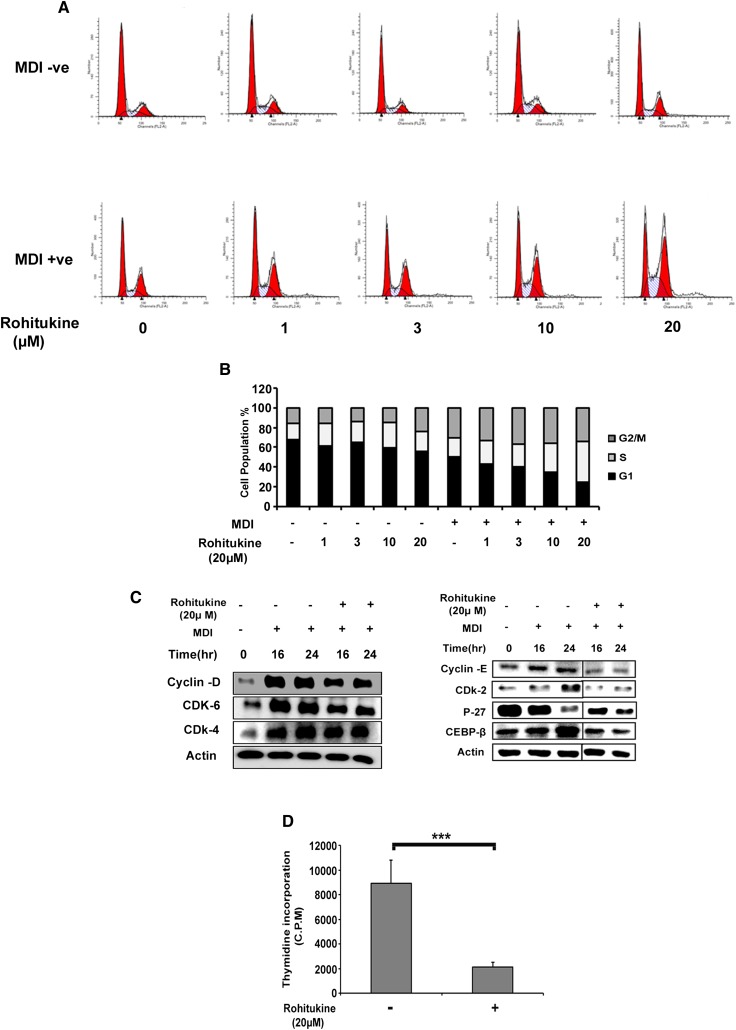
Fig. 4.
Effects of rohitukine on MDI-induced cell cycle progression in 3T3-L1 preadipocytes. A: Rohitukine arrested MDI-induced cell cycle progression in S phase. B: The population of cells in each stage of the cell cycle was quantified and showed concentration-dependent arrest in S phase. C: Postconfluent 3T3-L1 preadipocyte cells were incubated with MDI containing 20 μM rohitukine for 0, 16, and 24 h; rohitukine downregulated the expression of cyclin-D, CDK6, CDK4, cyclin-E, CDK2, C/EBPβ but stabilized P27 protein as observed by Western blotting. Represented cut blots were run, transferred, and exposed for the same time but separated away by unrelated samples. D: Rohitukine significantly inhibited the incorporation of [3H]thymidine into newly synthesized DNA after 48 h, measured by using a scintillation counter. The data are representative of three independent experiments that give similar results. Significance of differences between the MDI-induced control and rohitukine-treated groups: * P < 0.05, ** P < 0.01, *** P < 0.001.

